| Diese Seite auf Deutsch! |
|
Mussels and Clams (Bivalvia)Bivalvia Linnaeus 1758 (Acephala Cuvier 1798, Pelecypoda Goldfuss 1820, Lamellibranchia Blainville 1824) |
|
||||||||||||||||||||||||||||||||||
| Number of recent species in Mollusca, displayed by classes, including percentage. Sources: WoRMS: MolluscaBase eds. (2025): Mollusca Linnaeus, 1758. | ||||||||||||||||||||||||||||||||||
When walking along the beaches of Brittany in France, it is almost impossible not to notice the vast beds of blue mussels. Thousands of these dark bluish-black shells populate the coastal waters. At first glance, these mussel banks appear lifeless—nothing seems to move on their surface. But this impression is deceptive. A closer look reveals molluscs that have adapted in fascinating ways to a very special mode of life and feeding, and that play a crucial role within the coastal ecosystem.
At first sight, mussels appear very unlike other molluscs. Compared to a crawling snail - or even more so, to a squid darting through the water - they seem to have remained on a lower evolutionary level. Yet closer observation of a living mussel shows that this apparent immobility is in fact the result of a long evolutionary adaptation to a unique lifestyle.
Unlike all other molluscs, mussels usually are filter feeders. They draw not only oxygen but also food particles from the surrounding seawater. This method of feeding has proven so successful that mussels, over the course of evolution, have spread into an extraordinary variety of marine habitats, and even into freshwater environments.
The oldest known mussel and also the oldest known animal in general, was an Arctica islandica dredged off Iceland in 2016 and dated to be 507 years old.
![]() YouTube:
5 Most
Beautiful Clams In The World (ZoneA).
YouTube:
5 Most
Beautiful Clams In The World (ZoneA).
 Blue mussels (Mytilus edulis). Photo: Ron Offermans. |
A characteristic feature of mussels, visible from the outside, is their bivalved shell, which usually encloses the entire animal. The two shell valves are connected by an elastic hinge ligament, which, when relaxed, causes the shell to open. The antagonists of this mechanism are the powerful adductor muscles, with which the mussel can actively keep its shell tightly closed.
 Experiment with blue mussels: The glass on the left does not contain any mussels, the glass on the right shows how mussels filter the water. |
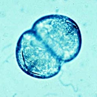
Most mussel species are dioecious (with separate sexes), though some groups are hermaphroditic. In addition, mussels that live in colonies (such as oysters, Ostrea) may undergo sex reversal, changing into males when too few males are present in the colony to ensure reproduction. While fertilisation and larval development in marine and many freshwater mussels take place in the open water, the large river mussels (Unionacea) carry out fertilisation and the early stages of larval development within the mantle cavity of the female. Their larvae, known as glochidia, develop through a parasitic stage by attaching themselves to a host fish. By contrast, most other mussel species develop through free-swimming planktonic larvae of the trochophore and veliger type.
 The interior of a mussel, schematic, one shell valve removes. Colour scheme cf. Body Plan of a Bivalve. Source: Biodidac, Modification: R. Nordsieck. |
 Model of a scallop (Pecten jacobaeus) from the Vienna Natural History Museum. Photo: Robert Nordsieck. |
The outer fold secretes the shell and the periostracum (the shell skin), the middle fold serves sensory functions, the inner fold regulates the water flow within the mantle cavity.
Due to the predominantly sedentary (sessile) lifestyle of most mussels, the head has regressed almost completely, except for the mouth region (for that reason, Cuvier in 1798 called the bivalves Acephala - headless molluscs).
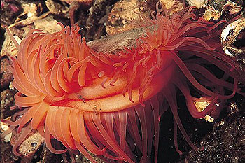
In swimming bivalves such as scallops (Pecten) and file clams (Lima), which require more detailed information about their environment, the mantle edge is lined with simple eyes (ocelli). In giant clams (Tridacna), symbiotic algae (zooxanthellae) live within the mantle tissue. The mussel provides protection for the algae, while in return benefiting from the photosynthetic products they produce.
![]() The Brain Maze:
The
bizarre visual system of scallops (Facebook Reel, AI Presentation).
The Brain Maze:
The
bizarre visual system of scallops (Facebook Reel, AI Presentation).
In burrowing or boring mussel species, the mantle openings are often extended into tube-like structures so that the animal can continue to draw in water and food while remaining buried in the substrate. These tubular extensions of the mantle are called siphons: an inhalant siphon for intake and an exhalant siphon for expulsion. Both may be fused into a retractable double tube, which in its extended state can even exceed the mussel's own length. The soft-shell clam (Mya arenaria), for example, lives buried in the sediments of the Wadden Sea and relies on its siphons to feed; if washed out of the sediment, it will die. By contrast, the blue mussel (Mytilus edulis) lives attached on the substrate and has no siphons: if it becomes covered by sediment, it too will perish.
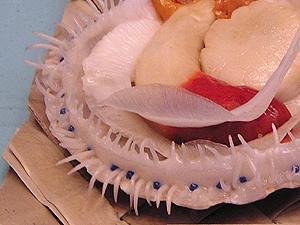
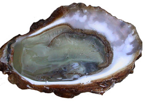 A freshly opened oyster (Ostrea edulis). |
The circulatory system of mussels, as in most molluscs, is open. The heart consists of two auricles and a single ventricle.
 Respiration and nutrition in a blue mussel. Source: Aquascope. |
 A clam digs itself into the ground using its foot. (see text). |
For humans, it is of particular concern that mussels tend to accumulate toxins in their tissues. The pollutants released by industry into the water ultimately end up in the blue mussels and oysters that we want to consume.
The mussel's foot can take on different shapes depending on lifestyle and locomotion; for example, beam-like, tongue-shaped, or worm-like. In swimming and sessile mussel species, the foot is often greatly reduced. In some species (such as the blue mussel Mytilus, the ark shell Arca, the scallop Pecten, or the pen shell Pinna), a byssus gland is located at the end of the foot. This gland secretes a proteinaceous substance that hardens in water into threads, allowing the mussel to anchor itself to the substrate. The byssal attachment can later be released when the mussel cuts off the threads (Mytilus) or sheds them entirely (pearl oyster Pinctada). Blue mussels also use their byssus threads defensively, entangling smaller snails such as dog whelks (Nassarius / Hinia).
 Swimming flame shell (Lima hians). Picture: Erling Svensen. |
Some bivalve groups (such as nut clams Nucula, bittersweet clams Glycymeris, tellins Tellina, and venus clams Venus) possess a true crawling foot, similar to that of snails.
Certain species (such as scallops Pecten and file clams Lima, see photo on the right) can even swim freely in the water. By snapping their two shell valves together rapidly, water is expelled from the mantle cavity, and the mussel is propelled in the opposite direction. Along the mantle edge of such mussels there are usually numerous simple eyes (ocelli), which provide information about light conditions in the environment, as well as tentacle-like appendages with which the mussel can sense its surroundings.
 A thick shelled river mussel's (Unio crassus) glochidia in a minnow's gills (Phoxinus phoxi- nus). Picture: Susanne Hochwald [2]. |
 Glochidium of a river mussel (U. crassus). Picture: Susanne Hochwald [1] |
![]() Glochidia: Larvae of Freshwater Mussels.
Glochidia: Larvae of Freshwater Mussels.
By contrast, most small freshwater mussels (pea clams Pisidium and fingernail clams Sphaerium) are hermaphrodites and give birth to living larvae (ovoviviparity). The zebra mussel (Dreissena polymorpha), which also belongs to the Dreissenidae, develops through a planktonic larval stage similar to the veliger, just like its marine relatives.
The systematic name of the bivalves (Bivalvia - the two-shelled molluscs) refers to their most characteristic feature: their shell, consisting of two separate valves. Adapted to the mussel's way of life, the shell varies greatly in form and serves as the most important characteristic for identification. It can be oval, elliptical, wedge-shaped, or sheath-shaped. The two valves may be very similar, especially in upright-living mussels such as river mussels, or very different in shape, especially in sideways-living species such as oysters (Ostrea) or scallops (Pecten).
The two valves are joined posteriorly by an elastic ligament, which opens the shell when relaxed. The mussel must actively close the valves against the ligament's resistance using one or two adductor muscles, which can be clearly seen when the shell is open. The inner dorsal margin of the valves is often thickened and may bear interlocking teeth that provide lateral stability and prevent slipping. The form of this hinge apparatus varies among mussel groups and is an important distinguishing feature.
 Hinge and ligament of a river mussel (Unio tumidus). Photo: M. Kohl. |
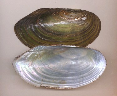 Shell halves of the swan mussel (Anodonta cygnea). Photo: M. Kohl. |
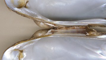
Most of the shell is composed of layers of prismatic crystals of the mineral
aragonite. This shell layer, the ostracum, may be covered externally by an
organic layer known as the periostracum. In addition, many mussels produce an
inner shell layer of plate-like aragonite crystals. This thin layer reflects
light in many colours (iridescence). If the mussel coats a foreign body trapped
between mantle and shell with this material, a pearl may form. For this reason,
the inner layer is also known as mother-of-pearl (hypostracum). Pearl mussels
exist both in the sea (e.g. Pinctada) and in freshwater (e.g.
Margaritifera in central Europe
and North America,
Hyriopsis and Cristaria
in East Asia). Human use of pearls in jewellery has a long history, with marine
pearls being far more common than the much rarer freshwater pearls.
Where
naturally formed pearls are insufficient to meet demand, pearls are now
cultivated by artificially implanting foreign bodies into mussels. Pearl
mussels, like blue mussels, are farmed in hydrocultures. Mussel farming,
especially of oysters and blue mussels, is now practised on almost all coasts of
the world.
The harvesting of pearls from the freshwater pearl mussel (Margaritifera margaritifera) has proven ecologically disastrous and, in the case of European populations, almost fatal. Today, the freshwater pearl mussel has become virtually extinct in rivers and streams, not only because of increasing water pollution but as a consequence of rampant uncontrolled pearl poaching over centuries. Moreover, its reproduction via glochidia depends exclusively on the brown trout (Salmo trutta fario), which has increasingly been displaced by the introduced rainbow trout (Oncorhynchus mykiss), an unsuitable host.
Not only because of their pearls, mussels are among the most economically important molluscs for humans. Prehistoric shell middens (Køkkenmøddinger, Danish for "kitchen waste heaps") have been found at human settlement sites and bear witness to the importance of mussels (mainly cockles and blue mussels) as a food source since the Palaeolithic.
Latest Change: 22.06.2026 (Robert Nordsieck).
