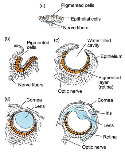
Stages in the evolution of eyes among molluscs. Source: Wikipedia.
a: Flat eye; b: cup eye; c: pinhole eye; d: vesicular eye; e: lens eye.
|
Stages in the evolution of eyes among molluscs. Source: Wikipedia. a: Flat eye; b: cup eye; c: pinhole eye; d: vesicular eye; e: lens eye. |
The example of the molluscs offers a good opportunity to observe the evolution of light sense organs in the animal kingdom. Among the numerous and various groups of molluscs there are primordial and advanced, movable and sessile species.
In the most primitive form light perception happens by single sense cells located somewhere in the body. Singular light sense cells dispersed over the body surface, as on snails and segmented worms, can tell the difference between light and dark, so the animal may benefit from a shadow reflex to protect itself against predators. They are, however, not a sense organ in the common sense of speaking: A sense organ is a complete organ, not just singular cells, specialized in a defined sensory performance. The first light sense organ is a specialized field of light sense cells and pigment cells for lateral isolation. It is called a flat eye. It enables its possessor to differentiate between light and dark, but only basically makes it possible to tell where the light comes from.
Flat eyes today can still be found in primitive groups of invertebrates, such as jellyfish (Coelenterata). It may also be assumed that the molluscs' ancestors, primitive, worm-like ground-living creatures, also possessed such flat eyes.
A primitive flat eye may be of valuable use to an animal either sessile or moving passively. The directed movement of more highly developed molluscs required the formation of more advanced light sense organs. In the consequence the light-sensitive epithelium of the flat eye caved in to form a pit. So the light sense cells on facing sides of the eye can tell apart light and shade. That makes it possible to determine where the light comes from. Pit shaped eyes can be found in sessile and slow moving invertebrates.
In adaptation to a directed movement there was not only an evolution of eyes, but also a change in body form: Sense organs became concentrated at the end of the body facing towards the the main direction of movement: The head evolved as the centre of sensory activity (cephalization).
While a pit eye may be able to differentiate between light and shade, it is not capable of producing pictures. Especially for predatory molluscs, having to observe and to follow their prey, an improvement of the eye's picture projection capability was necessary: The eye opening narrowed, and in consequence the picture projected on the retina became more focused. So the pigmented cup eye came into existence. Today, in its primitive state, this type of eye can be found among certain bivalves and turbellarian worms.
 A chiton. Source: Invertébrés Herbivores. |
Pigmented cup eyes can also be found among primitive, mainly sessile, gastropods, such as limpets (Patellidae).
Comparable to the pigmented cup eyes of primitive gastropods are the cuticular eyes of chitons (Polyplacophora). Those, as their name states, are situated in the dorsal shell plates of the chiton and enable the animal to tell apart light and shadow on its dorsal side.
In the further course of evolution, the eye opening reduced in size and as a result the eye achieved abilities comparable to a so-called pinhole camera: A focused, but low-light picture can be projected to the retina. Among the molluscs, pinhole eyes can be found among ormers (Haliotidae) and primitive cephalopods, such as Nautilus. Nautilus is a living fossil, a remnant from the Mesozoic. It is also assumed, that fossil cephalopods, such as the giant endocerate Cameraceras from the Ordovician had similar eyes.
![]() See: Nigel Marven's "Walking with dinosaurs: Sea Monsters"
See: Nigel Marven's "Walking with dinosaurs: Sea Monsters"
In the pit eye and the pinhole eye, the inner space of the eye is filled by a secretion breaking the light rays and, at least basically, enhancing brightness and focus of the picture. This inner eye space could evolve noticeably, when the eye opening of the pinhole eye closed completely and was covered by a translucent epithelium. Among more highly developed snails, especially carnivorous sea gastropods, this liquid-filled bubble inside the eye became a primitive lens, making possible the perception of a relatively focused picture with a usable brightness (in contrary to the pinhole eye, in which focus always works at the expense of brightness).
 A Roman snail's head. [CK] |
This vesicular or bubble eye reached its highest state of evolution among terrestrial snails: Looking at Roman snails (Helix pomatia) one can already discover a primitive lens eye. But there is no ciliar muscle in a snail's eye, which means the snail cannot focus the picture it sees by changing the lens, except by moving its tentacles, an ability that remains restricted to terrestrial snails, the terrestrial pulmonate snails (Stylommatophora), to be exact, among those also the Roman snail. Only they have got the eyes at the end of the long tentacles.
Eye stalks can also be found among some marine gastropods, for example conches (Strombidae). But they mainly enable the snail to look from under its shell without having to expose its head, not noticeably increasing its field of vision.
 A Roman snail's eye stalk. Picture: Martina Eleveld. |
Another method of focusing the picture can be found among bivalves (Bivalvia): Those do not have a head, but swimming species, such as scallops (Pecten) and flame shells (Lima), have relatively well evolved eyes in their mantle rim. A mirror tissue, the argentea, reflects the light, whose point of focus would be behind the retina, back on it and so produces a more focused picture.
The most highly evolved molluscs are cephalopods - cuttlefish, squids and octopuses. All of them are predators. Either they lurk and ambush their prey, or they hunt it actively. Their prey may be crabs or fish, but squids can compete in swimming even with the latter without effort.
To be able to achieve this, cephalopods need extremely good eyes: A Nautilus with its pinhole eye never would be able to compete with a fish, even without the cumbersome shell.
But squids have no shell (except a tiny inner shell, the gladius) and not only in swimming can compete with fish - also their eyes are more than adequate: They have got a lens, an iris and from outside look very much like a vertebrate eye.
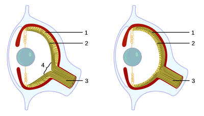 Inverse eye of a vertebrate (left) and everse eye of a mollusc (right). 1: Sense cells (retina); 2: nerve cells; 3: optical nerve; 4: blind spot. Source: Wikipedia. |
Only on a closer look one can see, that in the mollusc eye the sense cells point outwards, towards the light. During the development of the mollusc eye, the outer epithelium caves in and forms the eye ball. The light sense cells develop as a part of the eye wall. Only later are they connected to the nervous system. In contrary, the inverse vertebrate eye is formed first and invaded later by sensory cells and nerve cells from the brain. So the sensory cells point backward, to the eye wall.
 A cuttlefish's (Sepia officinalis) eye. Photo: Robert Patzner, Salzburg university. |
There is another joint character that, on a closer look, makes a cephalopod eye different from a vertebrate's. Both have ciliar muscles important for focusing the image. But in a vertebrate eye the ciliar muscle is used to deform the lens and so to adjust the point of focus. In a cephalopod eye, this happens by moving the lens forward and back and so changing the point of focus. The lens itself maintains its shape.
Their astonishingly good eyes enable cephalopods not only to compete with fish, but even to surpass them in some fields: There are remarkable methods of camouflage and communication in cephalopods. Octopuses can literally disappear in front of their background using colour cells (chromatophores) in their skin. Cuttlefish can communicate and transfer moods by changing colour. In some cuttlefish species, in mating time, there is an almost hypnotic play of rainbow colours.
Eyes under the microscope by Jan Parmentier.
Publication by the author: