| Diese Seite auf Deutsch! |
|
The Shell |
![]() See also:
The Evolution of the Mollusc Shell.
See also:
The Evolution of the Mollusc Shell.![]() Scalariform Snail Shells.
Scalariform Snail Shells.
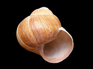 The shell of most Roman snails is coiled to the right: The shell mouth is placed on the right side of the snail's longitudinal axis. Photo: Robert Nordsieck. |
![]() The Way Snail Shells are Coiled.
The Way Snail Shells are Coiled.
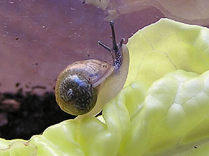 This juvenile Roman snail only has few whorls. Its shell is soft and translucent, so the internal organs can be seen. Photo: Robert Nordsieck. |
Chemically, the shell of a Roman snail consists mainly of lime (calcium carbonate). When the shell is placed in hydrochloric acid, the lime is decomposed with the release of carbon dioxide. However, an organic residue remains which is not attacked by the acid: while most of the shell is made of lime, it is externally covered by a very thin organic layer, the periostracum.
Under a microscope, a cross-section of the shell reveals several layers built of prism-shaped crystals of calcium carbonate. The crystals are arranged in successive, alternating directions, giving the shell great strength. This layer, which makes up by far the largest part of the shell wall, is therefore called the prismatic layer (ostracum). Bivalves and some marine gastropods possess an additional innermost shell layer, the hypostracum, which forms mother-of-pearl.
Snails cannot produce calcium by themselves – they must obtain it from their food or in other ways. On the one hand, calcium is absorbed from food during digestion; in addition, Roman snails can take up calcium directly from the soil when necessary. To do so, they remain at a calcium-rich spot and dissolve lime from the ground by acidifying their mucus with carbon dioxide, which is released through the sole of the foot.
The calcium obtained from food is stored mainly in the digestive gland (hepatopancreas) and, to a lesser extent, in the albumen gland. When required as building material for the shell, it can be transported in dissolved form, as calcium and carbonate ions, via the haemolymph to gland cells distributed over the entire surface of the mantle, which lies against the inside of the shell.
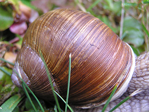 The growth stripes on this Roman snail's shell show one day's growth. Photo: Robert Nordsieck. |
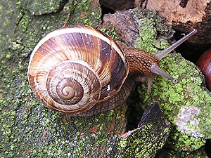 Juvenile Helix lucorum with well visible interruptions of shell bands because of growth breaks during periods of dryness. Photo: Robert Nordsieck. |
The growth lines visible on a Roman snail’s shell indicate how much the shell has grown in a single day. They can also be used to estimate the snail’s age at sexual maturity, when the formation of the reflected lip (see below) marks the end of shell growth. The spacing of the growth lines further reveals whether the snail had sufficient food and calcium supply at that time: when growth conditions are favourable, the shell grows faster and the lines are therefore wider apart.
 Growth of a (bivalve) mollusc's shell: A: Schematic longitudinal cut through the area of shell growth. B: Three-dimensio- nal view of wall growth. Prb: Ostracum growth; Pmb: Hypostracum growth. Ps: Pe- riostracum; Pr: Ostracum; Pm: Hypostracum; Ep: Pallial epithelium. d: dorsal; v: ventral. Source: Wehner, Gehring: Zoologie (1995). |
When a Roman snail reaches sexual maturity at about three years of age, the growth in size of its shell comes to an end. At the aperture, the shell margin develops a reflected lip that rounds off the previously sharp-edged opening. This reflected lip distinguishes the shell of an adult Roman snail from that of a juvenile.
The connection between sexual maturity and the completion of shell growth can be demonstrated by a pathological condition that occurs when a snail is infected by a parasitic fluke which destroys its hermaphroditic gland. As a result of this parasitic castration, shell growth continues unchecked — the affected snails can therefore be recognised externally by their abnormal gigantism. In such parasitically castrated Roman snails, no reflected lip is formed. A similar form of parasitic castration occurs in the mud snail (Galba truncatula) when it is infected by the common liver fluke (Fasciola hepatica).
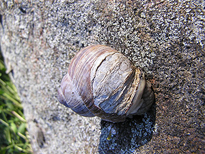 Roman snail (Helix pomatia) with a damaged and later repai- red shell. Photo: Robert Nordsieck. |
During their lifetime, most Roman snails sustain damage to their shells at some point, either through accidents or attacks by predators. Many of these snails, if they survive the incident, later show repaired shell areas that clearly differ from the rest of the shell surface. Although calcareous gland cells are distributed over the entire mantle surface and can produce material to repair the shell wall, a shell skin can only be formed by the specialised gland cells located along the outer edge of the mantle. Consequently, the subsequently repaired parts of the shell wall lack a shell skin - their granular, irregular surface structure distinguishes them from shell areas that have developed in the normal way.
A complete restoration of the entire shell wall, including the shell skin, is only possible while the shell is still growing. Once this growth has ended with the formation of the reflected lip at the aperture, only the shell wall can be reconstructed after damage, but not the shell skin itself.
Apart from shell damage caused by external influences, the shell of a Roman snail may also show other deformities, which, however, usually do not have any direct disadvantage for the animal.