| Diese Seite auf Deutsch! |
Torsion and the Coiled Snail Shell |
A snail's shell may be built following the same basic construction plan like a pond mussel or a nautilus, but its external appearance is quite different. A snail's shell is coiled to a spiral and the shell spire is visible on one body side only. Even in snails whose shell spire is not clearly visible, a closer examination will show the difference between apex (shell tip) on one side and umbilicus (shell navel) on the other.
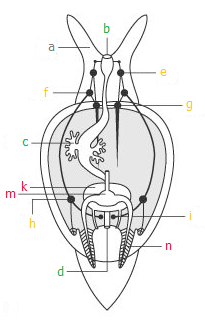 |
![]() Picture left: The basic state among
snails: a: Head tentacle. b - d:
Digestive tract: b: Mouth opening; c: Main digestive gland; d: Anus. e - i:
Nervous system: e: Cerebral ganglion; f: Pleural ganglion; g: Pedal ganglion; h: Parietal ganglion; i: Visceral ganglion. k - n:
Circulation: k: Heart bag
(Pericard); m: Heart; n: Pair of gills. Source: Kaestner,
A.: Lehrbuch der Speziellen Zoologie (Gustav Fischer, 1996), modified.
Picture left: The basic state among
snails: a: Head tentacle. b - d:
Digestive tract: b: Mouth opening; c: Main digestive gland; d: Anus. e - i:
Nervous system: e: Cerebral ganglion; f: Pleural ganglion; g: Pedal ganglion; h: Parietal ganglion; i: Visceral ganglion. k - n:
Circulation: k: Heart bag
(Pericard); m: Heart; n: Pair of gills. Source: Kaestner,
A.: Lehrbuch der Speziellen Zoologie (Gustav Fischer, 1996), modified.
The basis of this special evolution of the snail's shell lies in an evolutionary and embryonic process referred to as torsion (according to Haeckel's basic rule of recapitulation, evolutionary processes are repeated during embryonic development, which is why torsion took place during gastropod evolution and can also be observed during the embryonic development of the larva).
During torsion, the visceral sac with the mantle (responsible for building the shell) turns to the right, around its vertical (dorso-ventral) axis. While the most primordial gastropods had a cup-shaped bilaterally symmetrical shell, torsion took place only later, when the gastropods had already separated from the other molluscs.
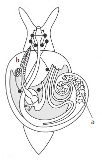 |
![]() Picture right: The state of torsion
among prosobranch snails: The visceral sac with the main digestive gland (a) has
moved to the right side of the body and the pallial cavity with the gills (b)
now is situated at the front. One gill has already been reduced. Source: Kaestner,
A.: Lehrbuch der Speziellen Zoologie (Gustav Fischer, 1996), modified.
Picture right: The state of torsion
among prosobranch snails: The visceral sac with the main digestive gland (a) has
moved to the right side of the body and the pallial cavity with the gills (b)
now is situated at the front. One gill has already been reduced. Source: Kaestner,
A.: Lehrbuch der Speziellen Zoologie (Gustav Fischer, 1996), modified.
As a consequence of torsion, the pallial cavity with the gills and the osphradium, a chemical-mechanical sense organ, moves to the front. Therefore, snail groups in this state of torsion are referred to as prosobranch snails (literally front-gill snails), a classification, that, however, cannot be maintained in systematics. It is assumed that originally advantage of torsion was a better water flow around the gills.
Because of the spatial changes the pallial cavity became narrower, so that one of the two gills was reduced during the consequent evolution. As a consequence, also one of the two atria of the heart was reduced, so since then snails' heart only have two chambers.
Neural pathways connect the original buccal ring of cerebral, pedal and pleural ganglia near the head and parietal and visceral ganglia in the body. Those neural pathways are called connectives. During torsion they were crossed, when the visceral sac was turning This state is referred to as chiastoneury or streptoneury, meaning crossed-nerve-state (from the Greek χιασμα = the cross and στρεπτος = coiled).
 |
Among opisthobranch snails (Opisthobranchia) a reverse evolution took place later. This Detorsion had the effect that the pallial cavity was situated first on the right side of the body (a state today still found in the group of side gill slugs (Notaspidea)) and finally came to rest at the back side of the body. During this process, the nervous system de-crossed again, so the connectives run straight again, a state called euthyneury (from the Greek ενθνς = straight).
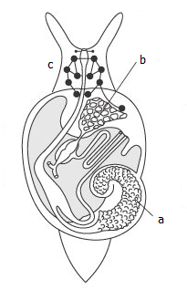
![]() Picture left: The development of
opisthobranch snails (Opisthobranchia). The pallial cavity (b) wanders
back to the back passing the right side of the body. The neural pathways
de-cross
(Euthyneury). a: Main digestive gland. Source: Kaestner,
A.: Lehrbuch der Speziellen Zoologie (Gustav Fischer, 1996), modified.
Picture left: The development of
opisthobranch snails (Opisthobranchia). The pallial cavity (b) wanders
back to the back passing the right side of the body. The neural pathways
de-cross
(Euthyneury). a: Main digestive gland. Source: Kaestner,
A.: Lehrbuch der Speziellen Zoologie (Gustav Fischer, 1996), modified.
In many opisthobranch snail groups the pallial cavity with the original gill has been reduced. Respiration now takes place using secondary gills on the snail's back, the cerata.
 |
The straight-nerve state (Euthyneury) is common to opisthobranch snails and pulmonate snails (Pulmonata). But the reasons are different: In pulmonate snails' evolution the ganglia had to a larger extent concentrated near the head (this ould be called the beginning of brain development or cerebralisation), so there never was streptoneury. Euthyneury in pulmonates has to be seen independent from that of opisthobranchs, though else there are many evolutionary traits joining both groups.
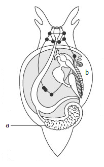
![]() Picture right: The evolution of
pulmonate snails (Pulmonata). While the pallial cavity with the lung (b)
remains in the front, like in prosobranch snails, there was no streptoneury,
because the ganglia were already concentrated near the head (c) and were not
influenced by torsion. a: Main digestive gland. Source: Kaestner,
A.: Lehrbuch der Speziellen Zoologie (Gustav Fischer, 1996), modified.
Picture right: The evolution of
pulmonate snails (Pulmonata). While the pallial cavity with the lung (b)
remains in the front, like in prosobranch snails, there was no streptoneury,
because the ganglia were already concentrated near the head (c) and were not
influenced by torsion. a: Main digestive gland. Source: Kaestner,
A.: Lehrbuch der Speziellen Zoologie (Gustav Fischer, 1996), modified.
For spatial reasons during torsion the visceral sac, together with the mantle and consequently also the shell were coiled to a spiral. Because at this point of evolution the visceral sac had already turned to one side of the body, the shell also is coiled asymmetrically to one side. Snails with a cup-like shell, like marine limpets (Patellidae) and fresh water limpets, acquired their special shell form only later. In the larvae, sometimes also in juvenile limpets, a shell spiral can be recognized.
As a summary, torsion can be understood as an advantageous development concerning the water flow into the pallial cavity. On the other hand, it made possible the reduction of paired organs such as the gills. The spirally coiled snail shell is unique among molluscs and in its special form has evolved only among gastropods. So snail shells can usually easily be recognised from shells of other mollusc classes.