The nervous system of snails, like that of all molluscs, is basically different from vertebrate nervous systems. Molluscs, with the exception of the most highly developed cephalopods, have no brain in the strict sense of the word. Instead, the cell bodies (pericarya) of nerve cells are concentrated in nerve knots (ganglia) in important parts of the body. Mollusc nerve cells have no myelin sheath. Therefore there can be no saltatory conduction. Instead, in the highly developed cephalopods, such as squids, giant axons have developed, whose large diameters make a faster transport of impulses possible.
Basically, the nervous system of molluscs can be derived from a rope-ladder nervous system, similar to that of segmented worms (Annelida). It is assumed that molluscs and annelids at least had a common ancestor. The mollusc nervous system, like that of annelids, is situated at the belly side (ventral side) of the animal (which is why they are called Gastroneuralia), and at least in primordial molluscs, remains of a metamerous (segmented) division of the nervous system can be recognised.
|
|
In gastropods, the ganglia originally have been dispersed over the body. As a remainder of the rope ladder nervous system, most ganglia come in pairs. Ganglia of the same type are connected by lateral connections, called commissurae. Ganglia of different types are connected by longitudinal connections called connectives. The most important ganglion types are the cerebral ganglia of the head, the pedal ganglia of the foot, the pleural and parietal ganglia of the pallial cavity and the visceral ganglia of the inner organs. In front of the cerebral ganglia there are the buccal ganglia of the pharynx.
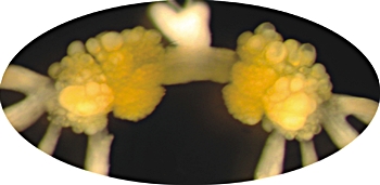
![]() Picture left: Oesophageal ganglia (buccal
ganglia)
of the sea hare Aplysia californica.
Source: Proekt,
A.; Wong, J.; Zhurov,
Y.; Kozlova, N.; Weiss,
K. R.; Brezina, V. (2008) "Predicting
Adaptive Behavior in the Environment from Central Nervous System Dynamics". PLoS
ONE 3 (11): e3678.
Picture left: Oesophageal ganglia (buccal
ganglia)
of the sea hare Aplysia californica.
Source: Proekt,
A.; Wong, J.; Zhurov,
Y.; Kozlova, N.; Weiss,
K. R.; Brezina, V. (2008) "Predicting
Adaptive Behavior in the Environment from Central Nervous System Dynamics". PLoS
ONE 3 (11): e3678.
 |
In most modern gastropod groups cerebral, pedal and pleural ganglia are concentrated and arranged together in a buccal ring surrounding the oesophagus near the head: The cerebral ganglia and pleural ganglia are situated above the oesophagus and the pedal ganglia beneath it.
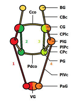
In marine gastropods, the visceral and parietal ganglia still are located in their original position in the body, connected to the buccal ring by long connectives. The mollusc nervous system is referred to as a tetraneural nervous system, because there are four main neural strands: Two pairs of connectives link the cerebral ganglia to the pedal ganglia on the ventral side. another to the visceral ganglia and parietal ganglia passing the pleural ganglia on the dorsal side.
![]() Picture right: Tetraneural nervous
system of gastropods: 1. and 4. nerve strand:
Cerebral ganglion (CG) - Cerebro-pleural connective (CPlc) - Pleural ganglion (PlG)
- Pleuro-visceral connective (PlVc) - Parietal ganglion (PaG) - Visceral ganglion
(VG). 2. and 3. nerve strand: Cerebral ganglion - Cerebro-pedal
connective (CPc) - Pedal ganglion (PG). Further ganglia and nerves:
BG: buccal ganglion.CBc: Cerebro-buccal connective. Cco: Cerebral commissura. PlPc: Pleuro-pedal connective.
Pdco: Pedal commissura. [RN]
Picture right: Tetraneural nervous
system of gastropods: 1. and 4. nerve strand:
Cerebral ganglion (CG) - Cerebro-pleural connective (CPlc) - Pleural ganglion (PlG)
- Pleuro-visceral connective (PlVc) - Parietal ganglion (PaG) - Visceral ganglion
(VG). 2. and 3. nerve strand: Cerebral ganglion - Cerebro-pedal
connective (CPc) - Pedal ganglion (PG). Further ganglia and nerves:
BG: buccal ganglion.CBc: Cerebro-buccal connective. Cco: Cerebral commissura. PlPc: Pleuro-pedal connective.
Pdco: Pedal commissura. [RN]
From the cerebral ganglia nerves lead towards the tentacles and the eyes, to the equilibrium organs (statocystes), to the lips and to the penis. The oesophageal buccal ganglia in front of them connect nerves to the gullet and the pharynx, the salivary glands and the stomach. The pedal ganglia innerve the foot muscle and the foot skin. The pleural ganglia are connected to the mantle. The parietal ganglia compute information from and to the gill and the osphradium, a chemo-mechanical sense organ in the pallial cavity. The visceral ganglia finally often are molten to one single ganglion and control nerves to the intestine, the anus, the skin, the posterior part of the genital apparatus, the kidney, the main digestive gland and the heart.
 |
During the so called torsion, during which process the visceral sac and the mantle turn around their vertical axis, so the pallial cavity originally located behind the visceral sac comes to lie in front of it, the visceral and parietal ganglia accompany them. After completion of torsion, the formerly right parietal ganglion now is on the left and the left on the right. Consequently, the formerly parallel connectives between the parietal ganglia and the buccal ring have been crossed: A situation called chiastoneury or streptoneury (crossed nerve state). Hence, gastropod groups, whose pallial cavity and gill are situated in front of the visceral sac are referred to as prosobranch snails (front gill snails) or Streptoneura.
 |
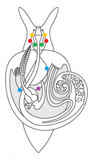
![]() Picture left:
Nervous system of a prosobranch snail. Yellow: Cerebral ganglia. Red: Pleural
ganglia. Green: Pedal ganglia. Blue: Parietal ganglia. Violet: Visceral ganglion.
The connectives between pleural and parietal ganglia are crossed as a
consequence of torsion (streptoneury). Source: Kaestner,
A.: Lehrbuch der Speziellen Zoologie (Gustav Fischer, 1996), modified.
Picture left:
Nervous system of a prosobranch snail. Yellow: Cerebral ganglia. Red: Pleural
ganglia. Green: Pedal ganglia. Blue: Parietal ganglia. Violet: Visceral ganglion.
The connectives between pleural and parietal ganglia are crossed as a
consequence of torsion (streptoneury). Source: Kaestner,
A.: Lehrbuch der Speziellen Zoologie (Gustav Fischer, 1996), modified.
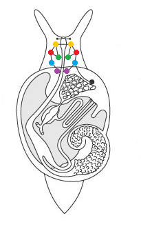
![]() Picture right: Nervous system of a
pulmonate snail (Pulmonata). Here the connectives between pleural and
parietal ganglia are not crossed (euthyneury), but the ganglia are concentrated
to such an extent that they have not been influenced by torsion. Source: Kaestner,
A.: Lehrbuch der Speziellen Zoologie (Gustav Fischer, 1996), modified.
Picture right: Nervous system of a
pulmonate snail (Pulmonata). Here the connectives between pleural and
parietal ganglia are not crossed (euthyneury), but the ganglia are concentrated
to such an extent that they have not been influenced by torsion. Source: Kaestner,
A.: Lehrbuch der Speziellen Zoologie (Gustav Fischer, 1996), modified.
During the further evolution of snails, escpecially connected to the transition to land, a concentration of all ganglia near the buccal mass took place. In pulmonate snails as well the pallial cavity is situated in front of the visceral sac. But in this group there is no streptoneury, because the ganglia had moved to far towards the head to be influenced by torsion.
Like in pulmonate snails, in the marine opisthobranch gastropods (Opisthobranchia) as well, there is no crossed-nerve-state. As a consequence, both groups had been joined as Euthyneura and opposed the Streptoneura, meaning the prosobranch snails,.
As many common traits pulmonate snails and opisthobranch snails may have, but in this regard they are distinctly different: While in pulmonate snails, as mentioned, the concentration of ganglia prevented streptoneury, in opisthobranch evolution it once was present, but later was reversed: During the so-called detorsion, the pallial cavity with the gills by a new right hand turn moved back behind the visceral sac and the neural connections again became parallel. So the reasons for euthyneury in pulmonates and opisthobranchs are distinctly different, albeit the result is the same.